In the May 19th online issue of PNAS, Dr. Che Alex Ma and colleagues presented the complete 3 dimensional model of the PBP1b protein on the surface of bacteria. This is the first time ever the mechanism of the enzyme that holds the key to bacterial cell wall formation is disclosed in details. The PBP1b contains 4 domains required for the synthesis of bacterial cell wall: the transglycosidase domain (TG), the transpeptidase domain (TP), the transmembrane domain (TM), and a yet to be defined domain called UB2H.
In the past 80 years, penicillin and its fellow antibiotic members have been used to fight off germ infections, what scientists really know was that these so called “antibiotics” are being effective by blocking certain key enzymes that are supposed to help germs construct their cell walls during the division and multiplication stage. These antibiotics target the cell-surface enzyme transpeptidase (TP). However, through the frequent use of these antibiotics, bacteria have become resistant to these drugs, so new antibiotics against the drug-resistant TP mutants or other new targets, such as TG, have to be developed to tackle the problem of antibiotic resistance. Now, 80 years later after penicillin was discovered, the Ma team from Genomics Research Center (GRC) of Academia Sinica in Taiwan reported the complete three dimensional picture of PBP1b in the advanced online edition in PNAS website this week.
According to Dr. Ma, a project he started 5 years ago when he came aboard GRC was to solve the mystery of cell wall synthesis. Well, this is definitely not a Rubic’s cube. First of all, one needs to have the thing in hand to study it. For this, his lab came up with a method to produce enough of the membrane-bound enzyme transglycosylase (TG), which can only be found on the cell surface of germs. He studied the activity of this enzyme and the work was published in early 2008.(See Prior Report)
The next big question is, just how exactly does TG do to conduct the task in making the cell walls? In their latest report, the research team coaxed this purified membrane protein into crystals and used a technology call x-ray crystallography to obtain its 3 dimensional structure. They laid out the clear picture of how TG takes an element called lipid II and keeps on performing kind of a knitting job, and finally makes the new skin when a germ divides into its own offspring cells. What’s more exciting is, the team has also identified several opportunities for future studies that may fill up the vacancies in finding cures for drug-resistant bacterial infections.
For the first time, the complete structure of the protein called PBP1b, with the multi-domain TG and a transmembrane (TM) domain can be seen clearly in a three dimensional way. In this model, they also revealed the interaction between moenomycin and TG. Moenomycin is an attractive studying target in the search of new antibiotics that functions by inactivating TG. Therefore, this is like showing a floor plan for those who are addressing the issue.
Furthermore, the team has identified an area within the whole structure so named as UB2H. With evidence shown, they proposed that UB2H is very well a key player in coordinating between the cell wall formation and DNA synthesis/repair. This may bring a crucial direction for future studies.
Lastly, the TM in the structure backs up strongly for the proposal of how cell wall can be synthesized at the membrane surface. The illustration can be seen in a Figure as well as an animation made by the team member.
“We did not envision so many achievements in one shot”, claimed Ma, “especially, each one turns on a light that we just cannot resist in digging along in the pursuit of facts which may eventually help fighting off bacterial infections.”
This research is part of a campaign to solve the drug resistance bacterial infection issue, led by President of Academia Sinica Dr. Chi-Huey Wong. Both Ma and Wong are the corresponding authors of this publication.
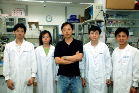 |
| Members: Ming-Ta Sung, Chia-Ying Huang, Che Ma, Lien-Yang Chou, Yen-Ting Lai (from left to right) |
It is also worth mentioning that, this is the first membrane protein structure that has ever been determined in Taiwan. Membrane proteins, corresponding to over 30% of all proteins, play critical roles in different biological processes and represent more than 50% of current drug targets. Of nearly 60,000 available structures of biological macromolecules in the Protein Data Bank (http://www.rcsb.org) now, only less than 1% are of membrane proteins.
Details of this publication can be found online in PNAS website: M.-T. Sung, Y.-T. Lai, C.-Y. Huang, L.-Y. Chou, H.-W. Shih, W.-C. Cheng, C.-H. Wong and C. Ma, “Crystal structure of the membrane-bound bifunctional transglycosylase PBP1b from Escherichia coli,” Proc. Nat. Acad. Sci. U.S.A., (2009).